Secondary glaucoma due to thrombosis of sigmoid and transverse sinus
Abstract
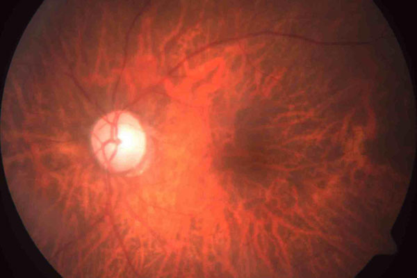
An 88-year-old female presented with redness in the left eye of one-month duration. On examination, the left eye showed 3 mm of proptosis with dilated and tortuous episcleral vessels and relative afferent pupillary defect. Intraocular pressure was 60 mmHg and showed open angles on gonioscopy with cup disc ratio of 0.8 in OS. A diagnosis of secondary open-angle glaucoma due to elevated episcleral venous pressure (EVP) was made. Magnetic resonance venogram revealed thrombosis of transverse and sigmoid sinus on the left side. This is the first case report of secondary open-angle glaucoma due to elevated EVP following thrombosis of transverse and sigmoid sinus.
References
Stamper RL, Lieberman MF, Drake MV (Eds). Becker-Shaffer’s Diagnosis and Therapy of the Glaucomas. 8th ed. St. Louis: Mosby; 2009:284.
Weinberg RN, Jeng S, Goldstick BJ. Glaucoma secondary to elevated episcleral venous pressure. In: Ritch R, Shields MB, Krupin T, eds. The Glaucomas. St. Louis: Mosby; 1989:1130.
Cioffi GA. Glaucoma: Basic and Clinical Science Course. San Francisco, CA: American Academy of Ophthalmology; 2014-2015:95.
De Keizer R. Carotid-cavernous and orbital arteriovenous fistulas: ocular features, diagnostic and hemodynamic considerations in relation to visual impairment and morbidity. Orbit. 2003;22(2):121-142.
Bousser MG, Ferro JM. Cerebral venous thrombosis: an update. Lancet Neurol. 2007;6:162-170.
Alvis-Miranda HR, Castellar-Leones SM, Alcala-Cerra G, Moscote-Salazar LR. Cerebral sinus venous thrombosis. J Neurosci Rural Pract. 2013 Oct-Dec;4(4):427-438.
Moses RA, Grodzki WJ. Mechanism of glaucoma secondary to increased venous pressure. Arch Ophthalmol. 1985;103:1653-1658.
Carvalho RA, Delloiagono HS, Jammal AA, Resende GM, Angotti HS. Secondary glaucoma following carotid cavernous fistula. Rev. Bras. Oftalmol. 2016;75(2):156-159.
Nassr MA, Morris CL, Netland PA, Karcioglu ZA. Intraocular pressure change in orbital disease. Surv Ophthalmol. 2009;54(5):519-544.
El Damarawy EA, El-Nekiedy AE, Fathi AM, Eissa ED. Role of magnetic resonance venography in evaluation of cerebral veins and sinuses occlusion. Alex J Med. 2012;48:29-34.

Copyright (c) 2020 Vijaya Pai H., Matta Rudhira Reddy

This work is licensed under a Creative Commons Attribution 4.0 International License.
Authors who publish with this journal agree to the following terms:
- Authors retain copyright and grant the journal right of first publication, with the work twelve (12) months after publication simultaneously licensed under a Creative Commons Attribution License that allows others to share the work with an acknowledgement of the work's authorship and initial publication in this journal.
- Authors are able to enter into separate, additional contractual arrangements for the non-exclusive distribution of the journal's published version of the work (e.g., post it to an institutional repository or publish it in a book), with an acknowledgement of its initial publication in this journal.
- Authors are permitted and encouraged to post their work online (e.g., in institutional repositories or on their website) prior to and during the submission process, as it can lead to productive exchanges, as well as earlier and greater citation of published work (See The Effect of Open Access).


