
Optimized imaging of the suprachoroidal space with swept-source OCT
Abstract
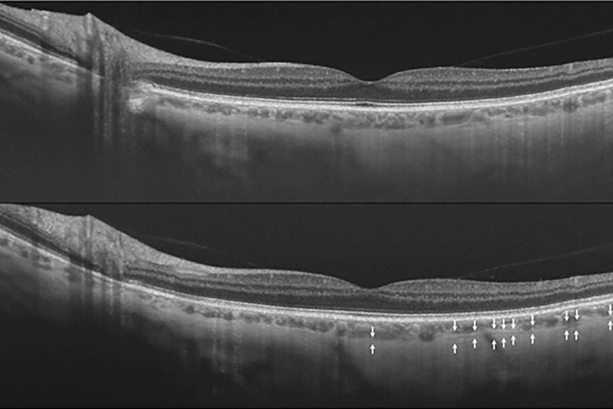
Purpose: To compare enhanced depth imaging (EDI) and non-EDI swept source optical coherence tomography (SS-OCT) in their ability to capture the suprachoroidal space (SCS).
Materials and methods: Twenty volunteers with a minimum age of 18 years without any ocular pathology and refractive error below Ã…} 2 diopters underwent SS-OCT foveal scanning, with and without EDI. Masked averaged B-scan lines were analyzed for presence of the SCS. When the SCS was seen, the percentage of the scan on which this structure could be unequivocally observed was measured. Scores obtained from the images taken with or without EDI were then compared.
Results: Thirty-seven eyes were analysed, since three eyes of three different patients were eliminated, as the outer border of the choroid was insufficiently delineated with both modalities. The SCS was not detected at all on 14 pictures (37.8%) obtained by non-EDI SS-OCT and 9 pictures (24.3%) obtained by EDI SS-OCT. When the SCS was detected with both modalities, it was observable on 27.2+/-24.2% of the scan without EDI and 40.4+/-30.3 of the scan with EDI (p < .001)
Conclusions: EDI SS-OCT enables a more frequent and extensive visualization of the suprachoroidal space than non-EDI SS-OCT. This new approach could be considered as the most accurate modality to currently visualize the SCS in vivo.
References
Moisseiev E, Loewenstein A, Yiu G. The suprachoroidal space: from potential space to a space with potential. Clin Ophthalmol. 2016;10:173-178.
Spaide RF, Koizumi H, Pozzoni MC. Enhanced depth imaging spectral-domain optical coherence tomography. Am J Ophthalmol. 2008;146(4):496-500.
Copete S, Flores-Moreno I, Montero JA, Duker JS, Ruiz-Moreno JM. Direct comparison of spectraldomain and swept-source OCT in the measurement of choroidal thickness in normal eyes. Br J Ophthalmol. 2014;98(3):334-338.
Adhi M, Liu JJ, Qavi AH, Grulkowski I, Fujimoto JG, Duker JS. Enhanced visualization of the choroido-scleral interface using swept-source OCT. Ophthalmic Surg Lasers Imaging Retina. 2013;44(6 Suppl):S40-S42.
Michalewska Z, Michalewski J, Nawrocka Z, Dulczewska-Cichecka K, Nawrocki J. Suprachoroidal layer and suprachoroidal space delineating the outer margin of the choroid in swept-source optical coherence tomography. Retina. 2015;35(2):244-249.
Yiu G, Pecen P, Sarin N, et al. Characterization of the choroid-scleral junction and suprachoroidal layer in healthy individuals on enhanced-depth imaging optical coherence tomography. JAMA Ophthalmol. 2014;132(2):174-181.
Kim JH, Chang YS, Kim JW, Lee TG, Lew YJ. Imaging suprachoroidal layer in exudative age-related macular degeneration. Curr Eye Res. 2015;41(5):715-720.
Boonarpha N, Zheng Y, Stangos AN, et al. Standardization of choroidal thickness measurements using enhanced depth imaging optical coherence tomography. Int J Ophthalmol. 2015;8(3):484-491.
Copyright (c) 2019 Joel Hanhart, Rozenman Yaacov

This work is licensed under a Creative Commons Attribution 4.0 International License.
Authors who publish with this journal agree to the following terms:
- Authors retain copyright and grant the journal right of first publication, with the work twelve (12) months after publication simultaneously licensed under a Creative Commons Attribution License that allows others to share the work with an acknowledgement of the work's authorship and initial publication in this journal.
- Authors are able to enter into separate, additional contractual arrangements for the non-exclusive distribution of the journal's published version of the work (e.g., post it to an institutional repository or publish it in a book), with an acknowledgement of its initial publication in this journal.
- Authors are permitted and encouraged to post their work online (e.g., in institutional repositories or on their website) prior to and during the submission process, as it can lead to productive exchanges, as well as earlier and greater citation of published work (See The Effect of Open Access).
